


위키페디아에서 가져온 그림과 설명이다.
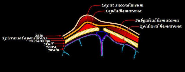
- Caput succedaneum is a neonatal condition involving a serosanguinous, subcutaneous, extraperiosteal fluid collection with poorly defined margins caused by the pressure of the presenting part of the scalp against the dilating cervix (tourniquet effect of the cervix) during delivery. It involves bleeding below the scalp and above the periosteum.
- A cephalohaematoma is a hemorrhage of blood between the skull and the periosteum of any age human, including a newborn baby secondary to rupture of blood vessels crossing the periosteum. Because the swelling is subperiosteal, its boundaries are limited by the individual bones, in contrast to a caput succedaneum.
- Subgaleal hemorrhage or hematoma is bleeding in the potential space between the skull periosteum and the scalp galea aponeurosis.
두피에서 일어날 수 있는 출혈(혈종)이나 부종에 대한 이해는 두피(머리뼈를 싸고 있는 피부를 포함하는 조직)에 대한 이해가 선생되어야 한다. 그런데 “두피”라는 것이 특별한 것은 아니다. 인체의 팔다리나 몸통에 있는 구조와 다르지 않다. 다만, 머리뼈를 덮고 있고 머리카락을 갖고 있다보니 얇게 존재하기 때문에 특별하게 보일 수 있지만, 결국 큰 줄거리에서는 인체의 다른 부위와 별반 다르지 않다. 그런 관점에서 두피를 본다면 그리 어려운 것은 아니다.
두피를 “scalp”라고 표현한다. 단순히 피부만을 뜻하는 것은 아니고, 피부 밑에 있는 근육과 결합조직층을 포괄적으로 표현한다. 따라서 실제 그 부위의 피부도 ‘두피’라고 표현하기 때문에 처음 책을 읽은 사람들 입장에서는 혼란스러울 수 밖에 없다.
참고로 나는 지금까지 머리 부분을 강의해 본 적은 없다.
그럼에도 불구하고 두피라고 특별히 공부를 해야하는 것은 아니다. 해부학구조라는 것은 큰 줄거리에서는 똑같기 때문이다. 학생들이 해부학을 어려워하는 이유도 부위마다 다른 구조라고 생각하고 접근하기 때문이라고 보여진다. 우리몸은 하나의 세포에서 발생하고, 발육하고 성장한다라는 큰 주제를 잊지 않으면 해부학이나 조직학, 발생학은 결코 어려운 학문이 아니다.
Scalp
scalp에 대한 위키페디아의 정의는 이렇다.
The scalp is usually described as having five layers, which can conveniently be remembered as a mnemonic:
- S: The skin on the head from which head hair grows. It contains numerous sebaceous glands and hair follicles.
- C: Connective tissue. A dense subcutaneous layer of fat and fibrous tissue that lies beneath the skin, containing the nerves and vessels of the scalp.
- A: The aponeurosis called epicranial aponeurosis (or galea aponeurotica) is the next layer. It is a tough layer of dense fibrous tissue which runs from the frontalis muscle anteriorly to the occipitalis posteriorly.
- L: The loose areolar connective tissue layer provides an easy plane of separation between the upper three layers and the pericranium. In scalping the scalp is torn off through this layer. It also provides a plane of access in craniofacial surgery and neurosurgery. This layer is sometimes referred to as the “danger zone” because of the ease by which infectious agents can spread through it to emissary veins which then drain into the cranium. The loose areolar tissue in this layer is made up of random collagen I bundles, collagen III. It will also be rich in glycosaminoglycans (GAGs) and will be constituted of more matrix than fibers. This layer allows the more superficial layers of the scalp to shift about in relation to the pericranium.
- P: The pericranium is the periosteum of the skull bones and provides nutrition to the bone and the capacity for repair. It may be lifted from the bone to allow removal of bone windows (craniotomy).
The clinically important layer is the aponeurosis. Scalp lacerations through this layer mean that the “anchoring” of the superficial layers is lost and gaping of the wound occurs which would require suturing. This can be achieved with simple or vertical mattress sutures using a non-absorbable material, which are subsequently removed at around days 7–10.
가장 쉽게 설명하자면,
팔다리가 되었던지, 몸통이 되었던지 간에 인체의 기본은 뼈이다. 즉, 뼈대계통(골격계통)이 중심이 되고, 그것을 움직일 수 있는 근육들이 붙는다(근육계통). 그리고 이것을 외부와 단절하고 보호하기 위해서 피부가 존재하는데, 근육위에 바로 피부가 있을 수 없기 때문에 그 사이에 결합조직들이 있게 된다(피하결합조직층). 이런 기본적인 구조를 머리에서도 똑같이 적용하면 된다. 다만, 머리뼈 위에 있는 피부를 포함한 결합조직은 다른 부위와는 달리 운동성이 없어도 되기 때문에(그렇다고 전혀 운동성이 없는 것이 아니다.) 그 부위의 근육층은 매우 얇을 것이라는 것을 염두에 두고 보면 쉽다. (그림은 모두 위키페디아에서 가져왔다.)
위 그림에서 보는 바와 같이 머리뼈(skull bone)을 싸고 있는 것은 인체의 다른 부위에서와 마찬가지로 골막(뼈막, periosteum)이다. 그리고 그 위의 모든 조직을 scalp(두피, 머리덮개)라고 한다. 그런데 앞서 위키페디아에 있는 “scalp”의 정의에서 SCALP 하나하나의 문자는 의미를 내포하고 있다는 것을 알 수 있다. 다시 간략하게 정리하면 아래와 같다.
- S: skin 피부
- C: Connective tissue 결합조직
- A: aponeurosis (or galea aponeurotica) 널힘줄(머리덮개널힘줄, 모상건막)
- L: loose areolar connective tissue layer 성근결합조직
- P: pericranium (periosteum) 머리뼈바깥막, 두개골막 (골막, 뼈막)
이 부분에서 혼란스러운 것이 있을 것이다. 바로 “pericranium, periosteum, 머리뼈바깥막, 두개골막, 골막, 뼈막)”이다. Scalp에 속한다고 했는데, 그 밑에 또 있으니 혼란스러울 것이다. 결론부터 말하자면 같은 구조물이다. 그런데 왜 저렇게 그림을 그려서 설명할까?
일반적으로 모든 뼈의 바깥층은 “periosteum (골막, 뼈막)”이다. 그런데 얇은 층으로 보이는 두피(scalp)의 마지막층인 “P”도 골막, 뼈막을 말한다. 그 층이 동일하다. 그림에서 각 층을 설명하면서 일반적으로 저렇게 그려놓기 때문이다.
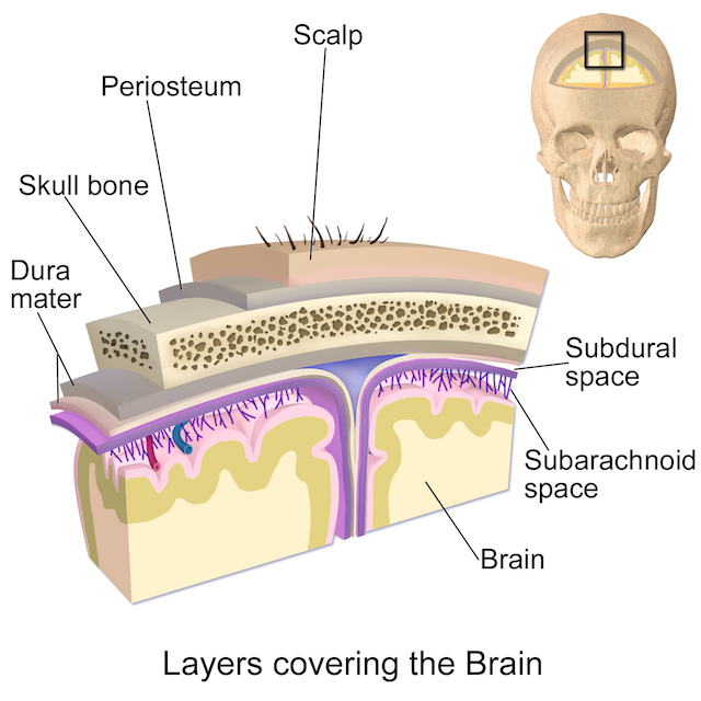
위키페이다에 나와 있는 아래 그림도 보면 좀 더 이해가 될 것이다. 머리카락을 갖고 있는 skin은 언급하지 않고 그아래 피부밑조직(피하조직)부터 표기를 하고있다. 그냥 피부는 일반적으로 알고 있다는 전제하에서 그려놓은 것이다. 따라서 해부학구조를 처음 접하는 학생들 입장에서는 혼란스러울 때가 있다. 그런데 항상 잊지말 것은 “인체를 덮고 있는 피부구조는 일정하다.”라는 대전제이다. 그것을 놓치면 안된다. 그리고 이 그림에서는 aponeurosis(gala aponeurosis)층과 pericranium(periosteum) 사이에 아무것도 그려놓지 않았다. 분명히 결합조직층이 있어야 하는데 말이다.
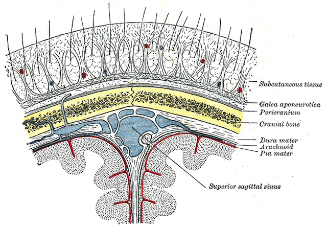
아래 그림도 마찬가지이다. 여기서는 피부밑조직(피하조직)을 노랗게 표시해두었고, aponeurosis는 얇게 흰색으로 표시해 두었다. 그리고 다행스럽게도 aponeurosis(gala aponeurosis)층과 pericranium(periosteum) 사이에 결합조직층(loose connective tissue)층을 노란색으로 잘 표시해 두었다. 자세히 보지 않으면 마치 pericranium(periosteum)으로 오해하게 그려두긴 했다.
굳이 이런 포스팅을 하는 이유가 이렇다. 이런 구조에 대한 기본적인 지식을 갖고 있는 사람이라면 이런 설명이나 그림이 아무런 문제가 되지 않는다. 그러나 처음 이런 구조물을 접하는 사람들에겐 어렵고 혼란스러울 수 밖에 없다. 특히 학생들은 더욱 그렇다. 나는 학생들에게 항상이야기한다.
인체는 하나의 세포에서 발생해서 발육하고, 성장한다. 따라서 이것을 하나의 거대한 한 개체로 이해하는데, 다만 부위별로 그 기능에 맞도록 특성화되어 있다는 것을 전체하고 접근하면 어려울 것이 하나도 없다.
나의 말
그럼에도 불구하고 해부학은 어려운 학문임에는 분명하다. 주일 아침 식사 중에 맨 위의 그림에 나와 있는 두피혈종에 대하여 아내에게 설명하면서 느낀 감정 때문에 이런 포스팅을 해둔다. 아내가 교과서로 보고있는 책은 설명이 틀렸다기 보다는 ‘학생들에게 혼란을 가져올 수 있는 책’이라는 생각이 들어서 이런 긴 포스팅을 하게 된 것이다.
사실 맨 위에 있는 위키페디아의 그림 설명도 매우 혼란스러운 그림이기도 하다. 엄밀하게 말하자면 잘못된 그림이다. 따라서 꼭 설명과 함께 보야야 한다는 것이다.
오전이 다 가고 있다. 발생학 시험문제 출제해야 하는데…. ㅠㅠ